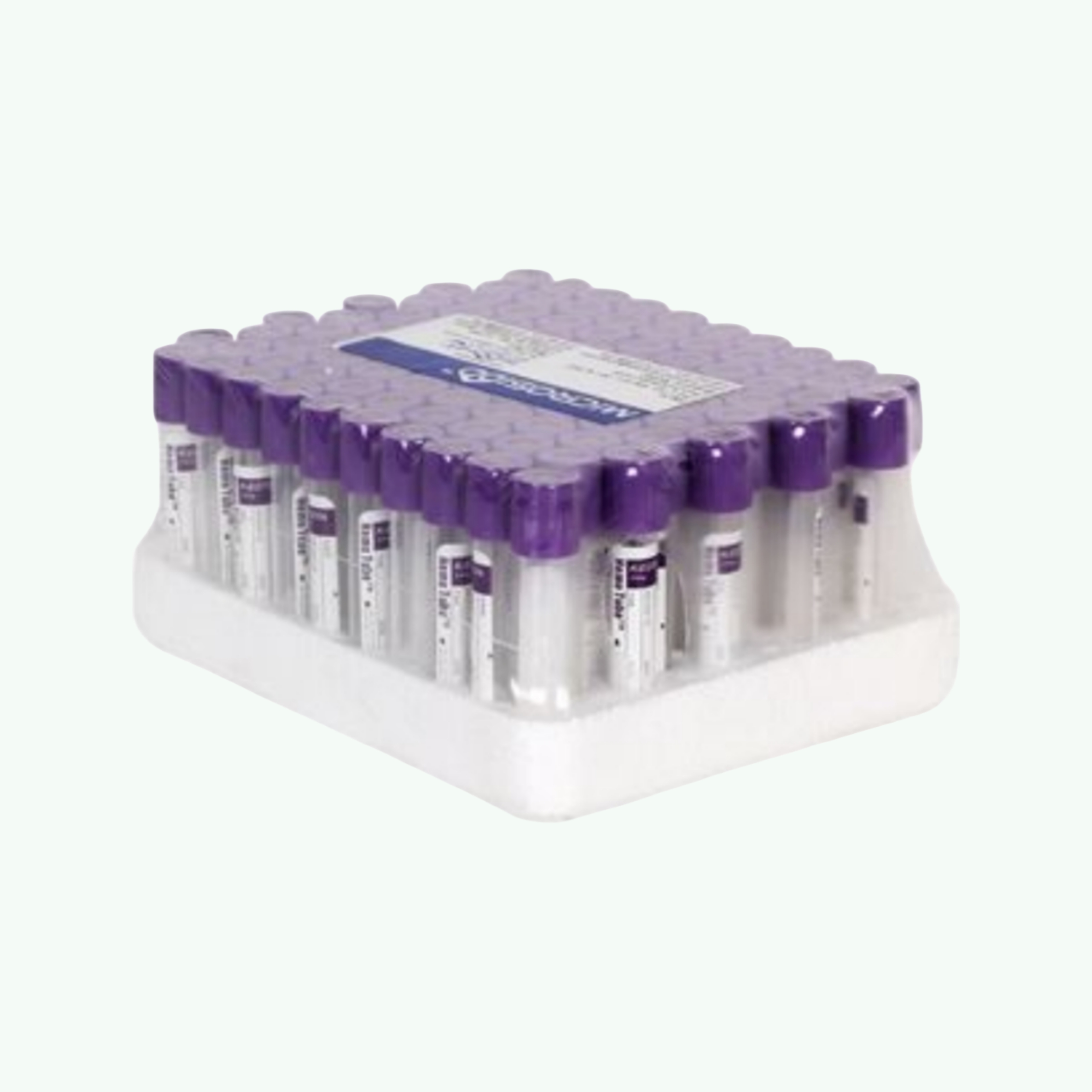
K3 EDTA Vacuum Blood Collection Tubes (Purple/Lavender Top)
Pack Sizes Available
Product Description
Technical Specifications
- Anticoagulant: K3 EDTA (tripotassium EDTA) — liquid solution form for rapid dissolution
- Cap/Stopper Colour: Lavender/Purple — internationally standardised colour code for EDTA
- Available Draw Volumes: 2ml, 3ml, 4ml, 5ml, 6ml (pre-evacuated to nominal draw volume)
- Tube Dimensions: 13 x 75mm (2-4ml) or 13 x 100mm (5-6ml)
- Pack Size: 100 tubes per box
- Primary Applications: CBC (haematology), blood film, blood group/cross-match, molecular testing (PCR/DNA)
Frequently asked questions
K2 EDTA (dipotassium EDTA) is typically supplied as a dry spray coating on the inner wall of the tube, while K3 EDTA (tripotassium EDTA) is supplied in liquid form as a solution in the tube. K3 EDTA dissolves more rapidly in the blood sample and provides faster, more reliable anticoagulation. However, K3 EDTA solution slightly dilutes the blood sample by the volume of the solution present, which can cause minimal lowering of erythrocyte counts and haematocrit in sensitive analyses. For most routine CBC testing, both perform equivalently. K2 EDTA is slightly preferred in CLSI/ICSH guidelines for haematology to avoid dilution, while K3 is preferred where faster dissolution is critical.
EDTA tubes are used for: complete blood count (CBC) including haemoglobin, haematocrit, white cell differential, platelet count and red cell indices; blood film morphology examination; reticulocyte count; erythrocyte sedimentation rate (ESR) in some protocols; blood group and cross-matching; and nucleic acid-based tests (PCR, molecular diagnostics) as EDTA preserves DNA and RNA integrity. EDTA is not suitable for coagulation studies (which require citrate tubes) or chemistry panels (which require gel separator or plain tubes).
After drawing the blood sample, the EDTA tube should be immediately inverted 8-10 times by gentle end-over-end rotation. This mixing ensures uniform distribution of the EDTA anticoagulant throughout the blood sample, preventing localised microclots that would affect the cell count. Do not shake the tube vigorously, as this can cause haemolysis and damage the cellular elements. Insufficient mixing is one of the most common pre-analytical errors in haematology and will cause platelet clumping and spuriously low platelet counts.
For routine haematology, EDTA blood samples should ideally be analysed within 4-6 hours of collection for optimal cellular morphology on blood films. Most automated cell count parameters remain stable at room temperature for up to 24-30 hours in K2 or K3 EDTA tubes. Refrigerated storage at 4-8°C extends stability to approximately 48-72 hours for cell count analysis. Blood films for morphology should be prepared as soon as possible after collection, as cellular changes at room temperature begin within a few hours.
K3 EDTA vacuum blood collection tubes with standard lavender caps and 13mm diameter tube bodies are compatible with the automated sample handling systems of most major haematology analyser brands including Sysmex, Beckman Coulter, Abbott, and Mindray platforms. The tube geometry (13x75mm or 13x100mm) should be confirmed against the analyser's sampling port specifications. For direct-draw or closed-tube sampling systems, the tube cap must be compatible with the needle penetration system of the specific analyser being used.
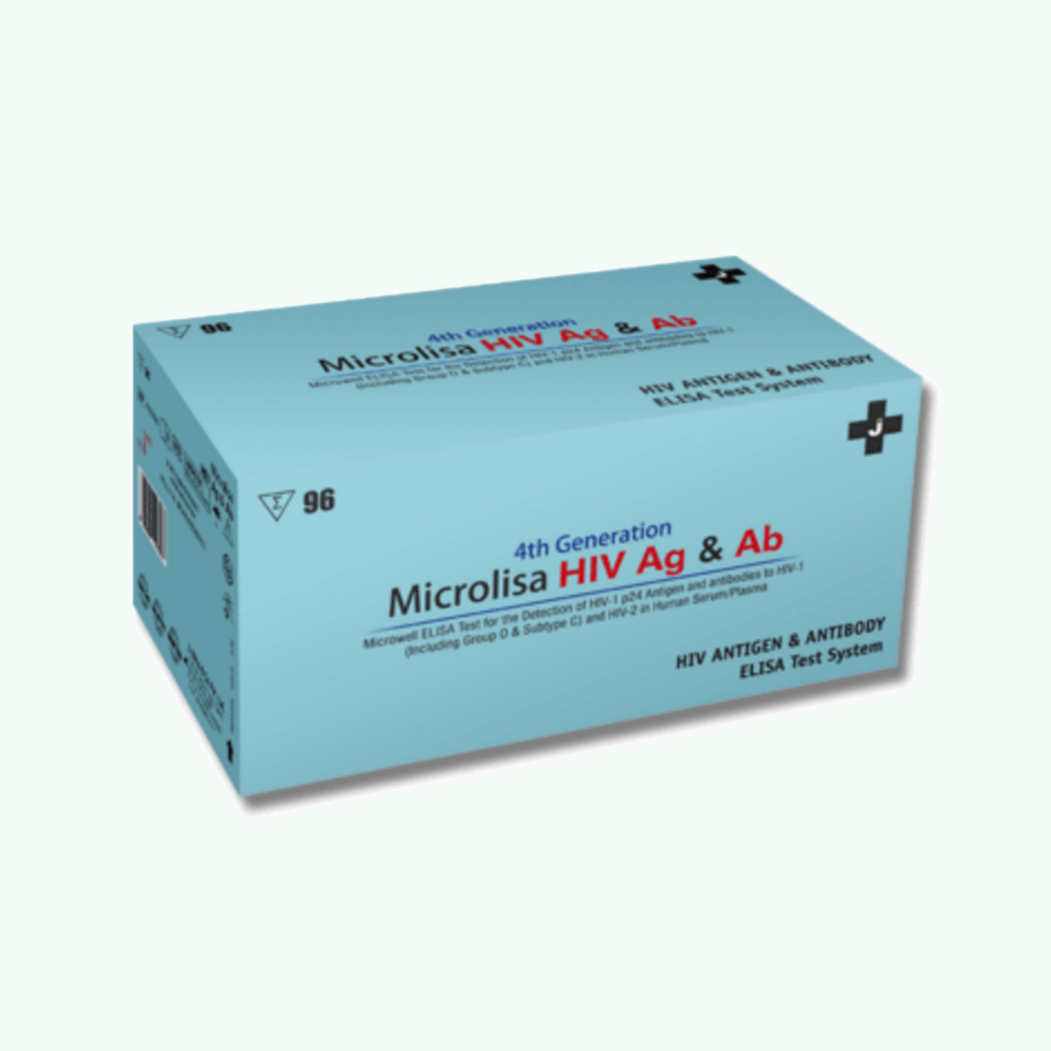
Microlisa HIV Ag & Ab 4th Generation ELISA Test Kit
Blood screening exists in a category of laboratory work where the margin for error is not just professionally unacceptable but medically catastrophic. A false negative in HIV screening does not just fail a test. It compromises patient safety, undermines transfusion protocols, and exposes healthcare systems to risks that nobody wants to calculate. Microlisa HIV Ag & Ab 4th Generation ELISA was engineered to close the detection window that makes early HIV infection so difficult to identify reliably. This is an in-vitro qualitative enzyme immunoassay designed for simultaneous detection of antibodies to HIV-1 (including Group O and subtype C prevalent in India), HIV-2, and HIV-1 p24 antigen in human serum or plasma. The test is intended for screening of blood donors, diagnostic testing of individuals at risk for HIV infection, and clinical evaluation of patients with AIDS-related symptoms. It represents the fourth generation of HIV ELISA technology, which detects both antibodies and antigens simultaneously rather than antibodies alone. The clinical advantage of 4th generation testing is the shortened window period. Traditional antibody-only tests miss early seroconversion cases where HIV-1 p24 antigen is present but antibodies have not yet developed to detectable levels. By detecting p24 antigen during the acute infection phase (typically 2 to 4 weeks post-exposure), this assay identifies infections approximately 1 to 2 weeks earlier than 3rd generation antibody-only tests. That earlier detection matters critically in blood donor screening and post-exposure monitoring. The assay is based on sandwich ELISA methodology. Microtiter wells are pre-coated with HIV envelope proteins (gp41, C-terminus of gp120 for HIV-1, and gp36 for HIV-2) and anti-p24 monoclonal antibodies. When specimens are added, any HIV antibodies or p24 antigen present bind to the coated antigens or antibodies. After washing, horseradish peroxidase (HRPO) conjugated antigens and anti-p24 antibodies are added, forming a sandwich complex. The colorimetric reaction develops proportionally to the amount of HIV antibodies or antigen present, read at 450nm absorbance. The kit uses color-coded reagents to monitor procedural steps, reducing protocol errors during multi-step workflows. Breakaway microwell strips allow testing flexibility from single specimens to full 96-well plate runs. Storage stability is maintained at 2-8°C with a shelf life of 24 months unopened. Total assay time including incubation steps is approximately 120 minutes. Sensitivity and specificity meet international standards for 4th generation HIV screening. Clinical evaluations demonstrate 100% sensitivity in detecting seroconversion panels and p24 antigen standards quantified down to 200 pg/ml. Specificity exceeds 99.5% when tested against large sample populations. The test detects all major HIV-1 subtypes including Group O and subtype C, which are epidemiologically significant in the Indian subcontinent. For distributors supplying blood banks, transfusion centers, diagnostic laboratories, and public health screening programs, Microlisa HIV Ag & Ab represents a clinically validated 4th generation screening platform with predictable reorder cycles. Sara Wellness has been exporting in-vitro diagnostic kits and laboratory reagents from India for 15 years.

Advantage PAN Malaria Card Rapid Diagnostic Test Kit
Malaria diagnosis in endemic regions operates under time pressure that microscopy cannot always accommodate. A patient presenting with fever in a rural health center at midnight does not have the luxury of waiting until morning for a trained microscopist to arrive, prepare slides, and spend twenty minutes examining blood films under oil immersion. That delay can mean the difference between timely artemisinin treatment and cerebral malaria developing overnight. Advantage PAN Malaria Card was designed to deliver species-level diagnosis in settings where microscopy is impractical or unavailable. This is a rapid visual immunoassay for qualitative detection of all four human Plasmodium species (P. falciparum, P. vivax, P. malariae, P. ovale) based on pan-specific plasmodium lactate dehydrogenase (pLDH) antigen in whole blood. The test provides results within 20 minutes using a simple fingerstick blood sample, no laboratory equipment required, making it ideal for point-of-care testing in primary health centers, rural clinics, field hospitals, and outbreak response settings. The assay is based on sandwich immunochromatography using monoclonal antibodies specific to pLDH, an enzyme produced by all Plasmodium species during their erythrocytic life cycle. When infected blood is added to the test device and assay buffer is applied, red blood cells lyse and pLDH antigen (if present) binds to gold-conjugated anti-pLDH antibodies. This complex migrates along the nitrocellulose membrane and is captured by immobilized anti-pLDH antibodies at the test line, producing a visible pink-purple band that confirms malaria infection. The see-through device design allows direct visualization of sample migration and result development, which helps identify invalid tests caused by insufficient sample volume or improper application. This transparency reduces the ambiguity that plagues some lateral flow devices where internal workings are hidden. Sensitivity and specificity have been validated through WHO malaria RDT evaluation programs using panels of wild and cultured parasites. The test detects parasitemia levels above 100 parasites per microliter of blood for both P. falciparum and P. vivax, which is clinically relevant for symptomatic infections requiring treatment. Specificity exceeds 99% when tested against cross-reactive conditions including dengue, leptospirosis, typhoid, and other febrile illnesses common in malaria-endemic areas. Shelf life is 24 to 30 months when stored at 4-30°C, which is critical for stockpiling in tropical climates where cold chain infrastructure is unreliable. The extended temperature stability means the test remains functional even when stored at ambient temperatures in resource-limited settings. Each kit contains individually sealed test devices, buffer vials, blood collection pipettes, and instructions for use. The test requires no special training beyond basic clinical skills and can be performed by nurses, paramedics, or trained community health workers. For distributors supplying national malaria control programs, public health departments, NGO field operations, and private diagnostic laboratories, Advantage PAN Malaria Card represents a WHO-evaluated rapid diagnostic platform with predictable consumption tied to malaria case loads. Sara Wellness has been exporting rapid diagnostic test kits and laboratory reagents from India for 15 years.
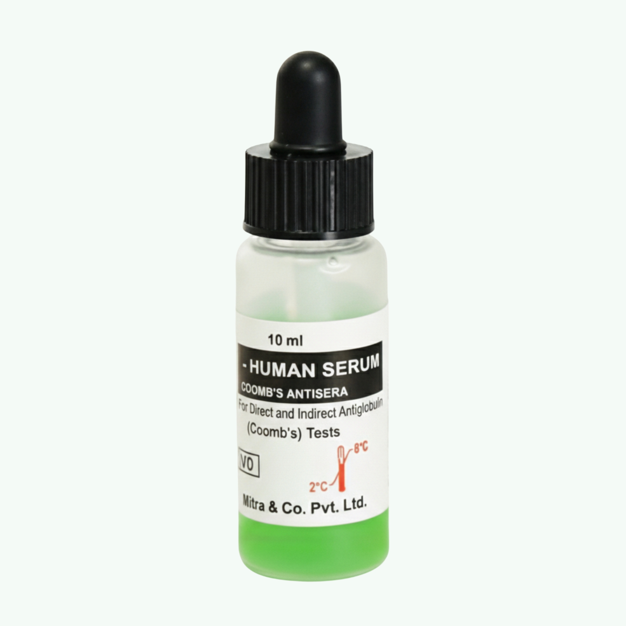
Human Serum Coombs Antisera (Antihuman Globulin Reagent)
Blood banking operates on a fundamental requirement that most people never think about until something goes wrong. Every unit of blood transfused must be confirmed compatible with the recipient's immune system. Every pregnant woman screened for antibodies that could harm her unborn child. Every suspected case of hemolytic anemia investigated for antibodies attacking the patient's own red blood cells. None of this happens without Coombs antisera making the invisible antibodies visible. Human Serum Coombs Antisera is the reagent that makes antiglobulin testing possible in blood banks and immunohematology laboratories worldwide. This is antihuman globulin (AHG) reagent used in both direct and indirect antiglobulin tests (Coombs tests) to detect antibodies and complement components bound to red blood cell surfaces or present free in serum. The reagent is produced by immunizing animals (typically rabbits) with human immunoglobulins, which induces production of polyclonal antibodies specific for human IgG antibodies and complement factor C3d. When added to washed red blood cells coated with IgG or complement, the antihuman antibodies bind to the human antibodies and form bridges between adjacent sensitized cells, causing visible agglutination. The direct antiglobulin test (DAT) detects antibodies or complement already bound to red blood cell surfaces in vivo. This test is critical for diagnosing autoimmune hemolytic anemia, investigating hemolytic transfusion reactions, and diagnosing hemolytic disease of the fetus and newborn. The indirect antiglobulin test (IAT) detects free antibodies circulating in serum or plasma. This test is essential for pre-transfusion antibody screening, crossmatching blood units for compatibility, and prenatal antibody screening in pregnant women. Polyspecific Coombs antisera (like the green-colored reagent shown) contains antibodies against both IgG and C3d complement, providing broad-spectrum detection. When the polyspecific reagent produces a positive result, monospecific antisera (anti-IgG alone or anti-C3d alone) are used for follow-up testing to characterize whether red cells are coated with IgG antibodies, complement, or both. This differentiation is clinically important because it helps determine the cause and clinical significance of the positive test. The reagent is typically dyed green using patent blue and tartrazine to allow easy visual identification during laboratory workflows where multiple reagents are used simultaneously. Storage at 2-8°C maintains reagent potency until the expiration date printed on the bottle, typically 18 to 24 months from manufacture. The reagent contains sodium azide (0.1% w/v) as a preservative, which inhibits bacterial growth but requires careful handling and disposal. Each dropper bottle delivers approximately 40 microliters per drop, allowing precise volumetric dosing during testing. The reagent must not be diluted and should not be used if turbid, as turbidity indicates bacterial contamination or protein aggregation that will compromise test performance. For distributors supplying blood banks, hospital transfusion services, reference immunohematology laboratories, and donor screening centers, Coombs antisera represents an essential reagent with consumption directly tied to transfusion volume and prenatal screening programs. Sara Wellness has been exporting immunohematology reagents and blood banking supplies from India for 15 years.